Before proceeding with clinical examination per se a good history taking is a must. Without proper history taking it is not possible to come to a reasonably correct diagnosis by clinical examination alone.
History should include:
Previous ear surgery
Previous head injury
systemic diseases like diabetes / hypertension
Use of ototoxic drugs
Exposure to noise during work
Family h/o deafness
H/O atopy / allergy
The classic symptoms of ear disease are as follows:
1. Deafness
2. Discharge
3. Tinnitus
4. Pain
5 Vertigo
Deafness: The patient must be asked whether deafness was sudden in onset, or gradual in onset. If deafness is sudden in onset the triggering event if any must be sought for. For example, deafness following head injury may be caused by a fracture of petrous portion of temporal bone. If the damage occurs to the auditory nerve the patient may have sensori neural hearing loss. Damage to 8th nerve is common following transverse fractures of temporal bone. Sometimes acute trauma may lead to dislocation of the ossicles causing conductive hearing loss. Of the 3 ossicles incus is the most commonly dislocated bone following trauma.
Conductive deafness can be differentiated from sensori neural deafness in a consious patient easily by doing a tuning fork test. Commonly used tuning fork tests are 1. Rinne, 2. Weber, and 3. Absolute bone conduction test.
Transient deafness after head injury may be commonly caused by a haematoma in the middle ear cavity. Following head injury the other common triggering event for deafness is viral infections. Common among them are mumps, measles etc. Deafness following viral infections are purely sensorineural in nature. The presence of wax is sufficient to cause fluctuating hearing loss which is conductive in nature.
Causes of fluctuating hearing loss are:
1. Presence of wax (conductive deafness) – Patient will c/o severe itching in the affected ear
2. Meniere’s disease (sensorineural deafness)
3. Peri lymph fistula (sensorineural deafness)
In patients with deafness associated with ear discharge the presence of perforation in the ear drum could be the cause.
In all patients with c/o deafness a proper drug history is a must. Ototoxic drugs like streptomycin, gentamycin and aspirin may cause irreversible damage to the hair cells of the cochlea causing sensori neural hearing loss. These drugs also sensitises the hair cells of the cochlea to damage due to noise exposure, hence occupational history of these patients is a must. H/O exposure to loud noise must be sought.
Discharge: Ear discharge is one of the common problems that brings the patient to the doctor. Before examining the patient a detailed history regarding
1. Duration of the discharge
2. Quantity of discharge
3. Quality of discharge
4. Aggravating & relieving factors must be sought for.
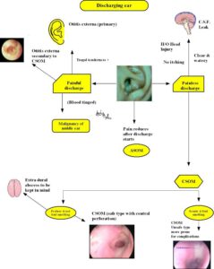
If the duration of discharge is short then acute conditions must be borne in mind. Common acute conditions which can lead to ear discharge are
1. A.S.O.M. – here the discharge is serosanguinous in nature (blood tinged), preceded by an episode of severe ear pain, pain subsides as soon as discharge starts, preceding epiosode of upper respiratory infection.
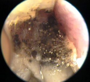
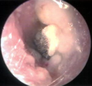
2. Otomycosis – common fungi affecting the external canal are candida and aspergillus fumigatus. Candida gives a curdy appearance in the external ear canal. In a dried up state it could appear like a cotton wool. Aspergillus fumigatus appears as a black color patches in the external auditory canal. These patients have ear discharge mostly just wetness, not profuse in nature, associated with intense itching.
3. C.S.F. Otorrhoea – The disharge is watery in nature, there is absolutely no mucoid elements in the discharge. This clear discharge starts when the affected ear assumes a dependent position. Biochemical analysis of this discharge will show that it contains glucose which is normally absent in purulent ear discharges.
Bedside test – One useful bedside test for CSF otorrhoea is Handkerchief test. If the secretion is mopped with a handkerchief and allowed to dry, there will be stiffening of the handkerchief if the discharge is from the middle ear because of the presence of mucous, if the discharge is csf there is no stiffening seen.
Most sensitive diagnostic test is estimation of Beta 2 transferrin in the secretions. Beta 2 transferrin is seen only in the CSF and is absent in other types of discharges.
Another important factor in the history taking is asking for the quantity of discharge. If the discharge is profuse the following conditions must be borne in mind : chronic mastoiditis, mastoid reservoir, extra dural abscess. Of these three in extra dural abscess the discharge is so profuse the external canal fills up with pus immediatly after mopping. The presence of mastoditis or mastoid reservoir can be ruled out by looking out for tenderness in the mastoid tip area. In children with well pneumatised mastoids the pus may cause erosion of the outer cortex and present as a collection just under the mastoid periosteum. This condition is known as sub periosteal abscess. If the ear discharge is scanty and foul smelling osteitic reaction due to infection must be suspected. This is frequently caused by the presence of cholesteatoma in the middle ear cavity associated with bone erosion.
The quality of discharge may range from:
Mucoid – common in CSOM
Mucopurulent – comon in CSOM associated with mastoiditis
Serous – Common in ASOM
Serosanguinous – ASOM and otitis externa
Watery – CSF otorrhoea
Tinnitus
Tinnitus is defined as hearing abnormal sounds in the ear. It can be classfied into objective tinnitus and subjective tinnitus. Objective tinnitus is the one which is heard by both the examiner and the patient eg palatal myoclonus. Subjective tinnitus is heard only by the patient. Even a simple problem like impacted wax can cause subjective tinnitus by the process of amplification of endogenous sound (internal mileu sounds of the body like the sound of circulating blood, contraction of muscle etc) Commonly tinnitus (subjective) in the absence of impacted cerumen indicates early sensori neural hearing loss. This is caused by damage to hair cells of the cochlea. The damage could be due to the adverse effects of medicines like those belonging to the group of antibiotics, diuretics or cytotoxic drugs. Tinnitus associated with hearing loss is commonly a manifestation of Menier’s syndrome. Tinnitus in this syndrome is roaring in nature and resolves within a day. It is also associated with giddiness.
Tinnitus in a patient with otosclerosis is an indication of active disease. These patients have active foci of otosclerosis. A separate term is used to identify these patients i.e. otospongiosis. Surgery if performed during this phase carries an immense risk of sensorineural hearing loss.
Pain:
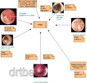
is one of the common complaints in patients with ear problem. Pain in the ear can arise from 2 sources, pain due to problems confined to the ear, and referred otalgia i.e. pain that is referred to the ear from a problem arising from other areas, i.e. pain associated with tonsillar infection has a propensity to radiate to the ear due to common nerve supply i.e. glossopharyngeal nerve. Pain due to inflammation in the external ear is intense and is associated with swelling of the external auditory canal. This can be differentiated from pain arising from middle ear inflammation by the presence of tenderness on pressing the tragus. This sign is known as the tragal sign. Tragal sign is negative in otalgia due to middle ear causes. Pain due to mastoiditis (inflammation of mastoid air cells) can be differentiated from pain due to otitis externa by the presence of three point tenderness. Three point tenderness is elicited by using the middle finger to apply pressure over the well of the concha, index finger is applied over the mastoid process, and the thumb is used over the mastoid tip. The pressure over the well of the concha indicates tenderness over the antral area, tenderness over the mastoid process indicates the presence of mastoiditis, and tenderness over the tip of the mastoid process indicate inflammation and thrombosis of mastoid emissary vein.
Vertigo: is defined as a sensation of unsteadiness / rotation. The commonest peripheral causes for vertigo are the diseases affecting the inner ear. It is always associated with tinnitus/ blocking sensation in the ear. Peripheral vertigo can be differentiated by central vertigo by its fatiguability. In peripheral vertigo the vertigo tends to diminish with time because the higher center learns to adjust with the problem. It is always positional. The patient will have to assume the provoking position for vertigo to manifest. Vertigo due to menier’s disease is self limiting and short lived. It never lasts for more than a day after which the patient gradually improves. Periperal vertigo is always associated with horizontal nystagmus, which is again fatiguing, where as central nystagmus due to cerebellar pathology manifests with rotatory / vertical nystagmus. They also show other postitive cerebellar signs like past pointing, dysdiadokokinesis etc.
Inspection:
The external ear is inspected with the following in mind:
1. size & shape of the pinna
2. Presence of tags / preauricular sinuses / pits
3. Evidence of trauma to pinna
4. Skin condition of pinna & external auditory canal
5. Evidence of previous surgery / presence of scars in the post aural / end aural region
6. Discharge from the external canal
7. Neoplastic lesions of pinna
The ear drum can be examined using an otoscope. The pinna should be grasped between the index finger and thumb and is pulled postero superiorly. This maneuver straightens the external canal bringing the ear drum into full view. This maneuver should be done only in adults. In infants the pinna must be pulled posteriorly and downwards in an effort to straighten the external canal. This is because of the fact the bony portion of the external canal is not fully developed in infants.
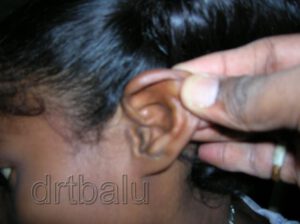
The use of Grubber’s aural speculum itself is sufficient to straighten the external canal. The status of the canal skin / presence or absence of discharge is noted. The whole of the ear drum is visualised by tilting and moving the otoscope in various directions.
The ear drum is roughly oval in shape and about 1 cm in diameter. Normal ear drum is pearly white in color. The following structures of ear drum are visualised:
1. Attic area
2. Pars tensa
3. Cone of light
4. Handle / lateral process of malleus
Rarely the following structures also can be seen:
Long process of incus
Head of stapes
Promontory
Eustachean tube orifice
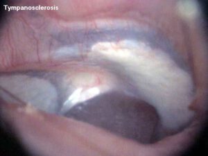
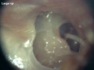
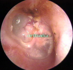
Perforations any must be identified, its position clearly documented. Through the perforation the status of the middle ear mucosa must be observed and documented. Presence of tympanosclerotic plaque (chalky mass over the ear drum) is an indicator of previous ear disease.
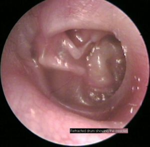
The cone of light must be observed for any distortion. Cone of light is absent in perforated ear drums, is distorted in retracted ear drums. It is also distorted when the ear drum is bulging as in the case of Acute otitis media.
The color of the ear drum must also be noted:
Red drum – is seen in acute otitis media, glomus jugulare
Blue drum – is seen in haemotympanum, secretory otitis media
Flamingo drum – is seen in otospongiosis
Mobility of the ear drum must be tested using a pneumatic otoscope, or a siegele’s speculum. The mobility of the ear drum is restricted in adhesive otitis media.
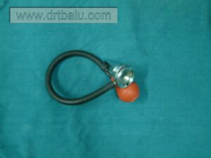
A siegel’s pneumatic speculum has an eye piece which has a magnification of 2.5 times. It is a convex lens. The eye piece is connected to a aural speculum. A bulb with a rubber tube is provided to insufflate air via the aural speculum.
The advantages of this aural speculum is that it provides a magnified view of the ear drum, the pressure of the external canal can be varied by pressing the bulb thereby the mobility of ear drum can be tested. Since it provides adequate suction effect, it can be used to suck out middle ear secretions in patients with CSOM. Ear drops can be applied into the middle ear by using this speculum. Ear is first filled with ear drops and a snugly fitting siegel’s speculum is applied to the external canal. Pressure in the external canal is varied by pressing and releasing the rubber bulb, this displaces the ear drops into the middle ear cavity.
Tests for hearing:
Useful bedside test for hearing is performed using a tuning fork. Ideally 3 frequencies are used 256 Hz, 512 Hz, and 1024 Hz. These three frequencies are used because they fall within speech frequency range. An ideal tuning fork should have the following features:
It should be made of a good alloy.
It should vibrate for one full minute.
It should not produce any over tones.
Tuning fork tests are performed to identify whether the patient is suffering from conductive deafness, sensorineural deafness, or mixed deafness. Three tests are performed towards this end. They are 1. Rinnes test, 2. webers test, 3. Absolute bone conduction test / ABC.
Rinnes test:
Ideally 512 tuning fork is used. It should be struck against the elbow or knee of the patient to vibrate. While striking care must be taken that the strike is made at the junction of the upper 1/3 and lower 2/3 of the fork. This is the maximum vibratory area of the tuning fork. It should not be struck against metallic object because it can cause overtones. As soon as the fork starts to vibrate it is placed at the mastoid process of the patient. The patient is advised to signal when he stops hearing the sound. As soon as the patient signals that he is unable to hear the fork anymore the vibrating fork is transferred immediatly just close to the external auditory canal and is held in such a way that the vibratory prongs vibrate parallel to the acoustic axis. In patients with normal hearing he should be able to hear the fork as soon as it is transferred to the front of the ear. This result is known as Positive rinne test. (Air conduction is better than bone conduction). In case of conductive deafness the patient will not be able to hear the fork as soon as it is transferred to the front of the ear (Bone conduction is better than air conduction). This is known as negative Rinne. It occurs in conductive deafness. This test is performed in both the ears.
If the patient is suffering from profound unilateral deafness then the sound will still be heard through the opposite ear this condition leads to a false positive rinne.

Weber’s test:
Here again 512 Hz tuning fork is used. The vibrating fork is placed over the forehead of the patient and he is asked to indicate on which side he is hearing the sound. Normally when hearing level is equal in both the ears, it is heard in the middle, in patients with conductive deafness the sound is heard in the left ear. This is known as lateralisation of Weber test. If the patient is suffering from sensorineural hearing loss then the sound is lateralised to the normal ear or the better ear. Hence weber’s test must always be interpreted along with the Rinne’s test. Weber’s test is a sensitive test, it can pin point even a 10 dB hearing difference between the ears.

Absolute bone conduction test:
This test is performed to identify sensorinerual hearing loss. In this test the hearing level of the patient is compared to that of the examiner. The examiner’s hearing is assumed to be normal. In this test the vibrating fork is placed over the mastoid process of the patient after occluding the external auditory canal. As soon as the patient indicates that he is unable to hear the sound anymore, the fork is transferred to the mastoid process of the examiner after occluding the external canal. In cases of normal hearing the examiner must not be able to hear the fork, but in cases of sensori neural hearing loss the examiner will be able to hear the sound, then the test is interpreted as ABC reduced. It is not reduced in cases with normal hearing.
Basic tests for hearing:
For making a basic assessment of patient’s hearing the ear opposite to the one tested is masked by occluding it. The patient is asked to repeat random numbers uttered by the examiner. Ideally patient is blind folded to prevent lip reading. The numbers are uttered at various intensities, quiet whisper, loud whisper, quite voice, loud voice and shout.
Rough estimation of hearing loss would be:
quite whisper – normal
Loud whisper – 20 – 30 dB
Quite voice – 30 – 45 dB
Loud voice – 45 – 60 dB
Shout – 60 – 80 dB