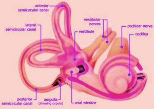
Benign paroxysmal positional vertigo is the most commonly diagnosed vestibular disorder. This is commonly caused by dysfunction of the posterior semicircular canal. Lateral and superior semicircular canals can also be involved on rare occasions. It is characterized by brief spells of severe vertigo (often lasting for just a few seconds) that are experienced only with specific movements of the head.
History:
This disorder was first described by Barany in 1921. He documented the various components of this disorder as 1. Nystagmus, 2. Fatiguability of the nystagmus and 3. Vertigo. He failed to correlate the onset of nystagmus with specific positions of the head.
Dix & Hallpike 1952 described the Dix Hall-pike maneuver for eliciting the nystagmus. They also described the unique features of nystagmus accompanying this disorder. These features were 1. Very short latency, 2. Directional features, 3. Brief duration, and 4. Reversibility on returning the patient to a seated position.
Schuknecht postulated th
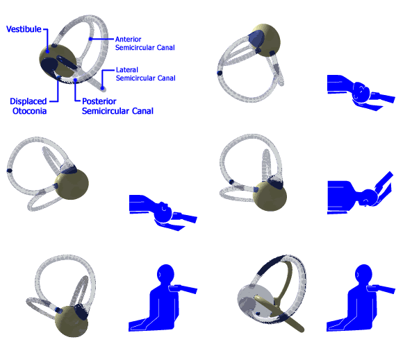
at BPPV was caused by loose otoconia from the utricle which in certain positions, displaced the cupula of the posterior canal. (Schuknecht theory). He later modified his theory and proposed that it was due to the deposition of otoconia on the cupula of the posterior semicircular canal. He termed this theory as cupulolithiasis. The cupulolithiasis theory proposes that calcium deposits become embedded on the cupula making the posterior semicircular canal sensitive to gravity.
Hall & Ruby suggested that BPPV could result from deflection of the posterior canal cupula caused by debris within the posterior canal. This theory became known as the canal lithiasis theory. In this theory the calcium debris doesnot become adherent to the cupula but float freely within the canal. Head movements like looking up, down, or rolling over to the affected ear may result in the displacement of the sludge causing the classic symptoms.
Hall & Ruby described 2 types of BPPV: 1. BPPV with a fatiguable nystagmus, where the deposits are freely mobile within the cupula of the posterior canal,
2. BPPV with a non fatiguing nystagmus where the calcium deposits are fixed on the cupula of the posterior canal.
Typical features of BPPV as described by Hall & Ruby:
1. Canalithiasis mechanism – This explains the latency of the nystagmus as a result of the time needed for motion of the material within the posterior canal to be initiated by the gravity.
2. Duration of the nystagmus – is correlated with the length of time required for the dense material to reach the lowest part of the posterior canal.
3. The vertical (upbeating) and torsional (superior poles of the eye beating towards the lowermost ear). The nystagmus is more vertical when the patient looks away from the lowermost ear, and more torsional when looking towards the lowermost ear.
4. The reversal of nystagmus when the patient returns to the sitting position is due to retrograde movement of material in the lumen of the posterior canal back towards the ampula, resulting in ampulo petal deflection of the cupula.
5. The fatiguability of the nystagmus evoked by repeated Dix Hallpike positional testing is explained by dispersion of material within the canal.

Incidence:
BPPV is the most common cause of vertigo constituting 20 – 40% of all patients with peripheral vestibular disease. Mean age of onset ranging between 4th and 5th decades. women outnumbering men by 2:1.
History: Patient c/o severe vertigo associated with change in head position. Symptoms are always sudden in nature, never lasting more than a minute. The patient may even volunteer provocating postures.
On examination: the classic eye movements associated with Dix Hallpike maneuver is seen.
Dix-Hallpike maneuver: The patient is positioned on the examination table in such a way that when he/she is placed supine, the head extends over the edge. The patient is lowered with the head supported and turned 45 degrees to one or the other side. The eyes are carefully observed; if no abnormal eye movements are seen, the patient is returned to the upright position.
This same maneuver is repeated with the head in the opposite direction and the patient’s symptoms are noted.
The pattern of response consists of the following:
1. Nystagmus is a combination of vertical upbeating & rotatory (torsional) beating towards the downward eye. Pure vertical nystagmus is not seen in BPPV.
2. There is often a latency of onset of nystagmus
3. Duration is less than a minute
4. Vertiginous symptoms are invariably seen
5. Nystagmus disappears with repeated testing (fatiguability)
6. Symptoms often recur with the nystagmus in opposite direction on return of the head to upright position.
Canalithiasis involving the posterior canal is the commonest cause of BPPV. Posterior canal BPPV may rarely be bilateral, but while testing the head must be positioned in the plane of the posterior canal during testing of unaffected ear otherwise the debris in the affected side can rest against the cupula and stimulate an exitatory nystagmus from the unaffected ear.
Lateral canal BPPV:
Lateral canal has also been identified as the offender in 17 % of cases with BPPV. Lateral canal BPPV can be detected by a variation of Dix Hallpike maneuver. The patient’s head is first brought to the supine position resting on the examination table (not hyperextended). The head is then turned rapidly to the right so that the patient’s right ear rests on the table. The eye movements of the patient are monitored with Frenzel’s glasses for 30 seconds. The patient’s head is then turned to the supine position (eyes looking upward) and is then rapidly turned to the left so that the left ear rests on the table. Eye movements are monitored. The nystagmus with lateral canal BPPV is horizontal and may beat toward (geotropic) or away (ageotropic) from the downward ear. It begins with a short latency, increases in magnitude progressively, and is less susceptible to fatigue with repetetive testing than the vertical torsional nystagmus of posterior canal BPPV.
Cupulolithiasis, either alone or in combination with canalithiasis is more likely to be involved in the etiology of lateral canal BPPV than in the case of posterior canal BPPV. If the nystagmus is geotropic, the particles are likely to be in the long arm of the lateral canal relatively far from the ampulla, if the nystagmus is ageotropic, the particles could be in the long arm relatively close to the ampulla or on the opposite side of the cupula either floating within the endolymph or embedded in the cupula.
Superior canal BPPV: Incidence of superior canal BPPV is very rare.
Standard electrooculography or 2 dimensional video nystagmography devices donot record the typical eye movements associated with BPPV. Thus clinical examination of the patient is of paramount importance.
Management:
Medical:
Repositioning maneuver: Currently BPPV is managed by repositioning maneuvers that, in cases of canalithiasis use gravity to move canalith debris out of the affected semcircular canal and into the vestibule. For posterior canal BPPV the manuver developed by Epley is effective.
Epley maneuver – This is performed by placing the head of the patient in the Dix Hallpike position that evokes the vertigo. The posterior canal on the affected side is in the earth vertical plane when the head is in this position. After the cessation of initial nystagmus, the head is rolled through 180 degrees, (this is done in two 90 degree increments, stopping in each position until the nystagmus resolves) to the position in which the offending ear is up. The patient is then brought to the upright sitting position. This procedure is likely to be successful when nystagmus of the same direction ccontinues to be elicited in each of the new position (as the debris continues to move away from the cupula). This manuver is repeated until no nystagmus is elicited. This is successful in 90 % of cases. Posterior canal BPPV can be converted to lateral canal BPPV during Epley maneuver. The lateral canal BPPV resolves in several days. Drugs are usually not prescribed, but low dose meclizine or calmpose ccan be given 1 hour before the procedure if the patient is anxious or prone to vomiting.
Sermont maneuver – is also effective in posterior canal BPPV, but is most difficult to perform and it has no significant advantages over the Epley maneuver. This is being described here for the sake of completion. In this maneuver the patient is moved quickly in to the position that provokes the vertigo and remains in that position for 4 minutes. The patient is then turned rapidly to the opposite side ear down, and remain in the second position for 4 minutes before slowly getting up.
In both these maneuvers gravity is the stimulus that move the particles within the canal, so there is no need to turn the head on the body, enbloc movement of the head and body as much as possible is the plan.

Image showing Re positioning maneuver
Vibrator therapy:
Some physicians use a small hand held vibrator over the mastoid to agitate the particles and make it move. This mastoid vibrator is to be avoided in patients with retinal detachment or in patients who may be susceptible to retinal detachement due to high myopia.
After these repositioning manuvers patients are instructed to avoid bending over and are told to sleep with the head elevated atleast 45 degreees for the next couple of days.
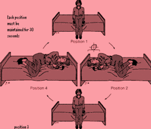
Brandt Doroff exercises – can be performed by the patient in the home environment. These exercises are performed in 3 sets / day for 2 weeks.
It is started like this:
Position 1 – The patient must be seated upright on the bed. Then he moves to side lying position (position 2) the head is kept angled upwards about half way. The patient should stay in this position at least for 30 seconds or till the giddiness subsides. If the giddiness does not subside thee patient must revert back to position 1. After 30 seconds the procedure is repeated on the opposite side. Most of the patients get relief within a period of 10 minutes.

Treatment manuvers for lateral canal BPPV:
In these patients with geotropic nystagmus lying on one side with the affected ear up for 12 hours has been found to be effective.
Surgical management:
Singular neurectomy – is a very demanding procedure. The posterior canal is supplied by singular branch of vestibular nerve. This nerve when preferentially sectioned alleviates the patient’s symptom due to posterior canal BPPV.
Posterior canal plugging procedure – is a easier procedure. Through a mastoidectomy incision the labyrinth is exposed. The posterior canal is drilled exposing the membranous portion of the canal. The canal is sealed and packed off thereby preventing the debris from floating. After the procedure the patient may feel slightly giddy. The patient needs to be kept in the hospital till giddiness subsides.